
Optical coherence tomography (OCT) or digital retinal imaging, takes photos of the retina, the light sensitive part in the back of your eye, quickly and non-invasively without the use of radiation or x-rays. It uses light to take high resolution 3D images of the important structures in your eye, including the optic nerve, blood vessels, and macula. It also produces a scan of the retina’s distinctive layers with near-cellular resolution. These detailed scans are a useful tool in closely monitoring your eye health over time.
OCT helps with early detection, management, and monitoring of many ocular conditions. Many common eye conditions may develop without any noticeable symptoms until they have progressed quite significantly. Many of these can worsen if left undiagnosed, and may lead to visual impairment or in some cases, complete vision loss. The earliest indications of conditions like macular degeneration, diabetic retinopathy, and glaucoma can be detected by an OCT.
Your OCT results will be reviewed with you during your eye exam. It’s a really great way to get a better understanding of your eye health because any issues can be seen clearly on the photos.
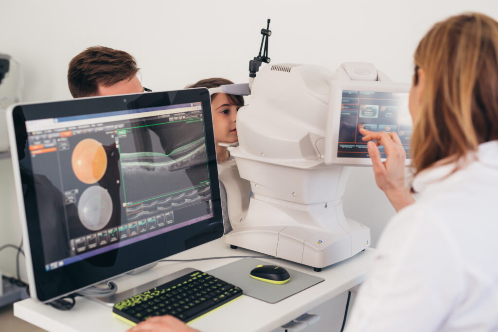
We recommend OCT imaging for everyone, including children. OCT imaging takes less than one minute and the process is painless, with nothing touching your actual eye.
OCT images are especially recommended for those with eye disease, risk factors for eye conditions, high prescriptions, or a family history of eye disease. We also recommend OCT imaging for patients who are light sensitive as we can more thoroughly analyze the anatomy of your eyes without the prolonged use of lights, allowing you to be more comfortable during your eye exam.
Even if you don’t have a high risk of eye conditions, OCT imaging can create a baseline to better detect the smallest changes to your eyes over time.
At Milton Optometry, OCT imaging is included in all non-OHIP insured annual eye exams. OCT imaging is not covered by OHIP but can be added to any exam and may even be covered by your private insurance. Bring your insurance card and we can direct bill your OCT.
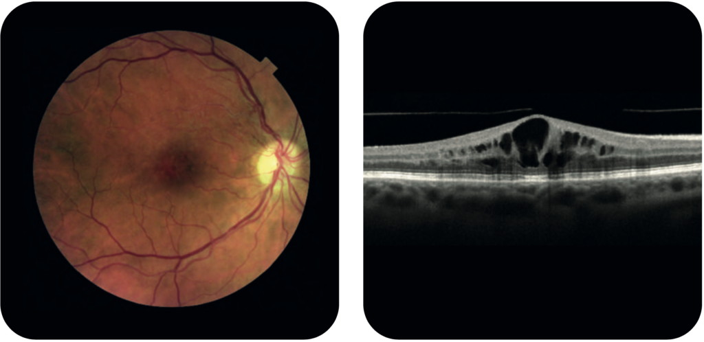
Normal retina and retinal scan
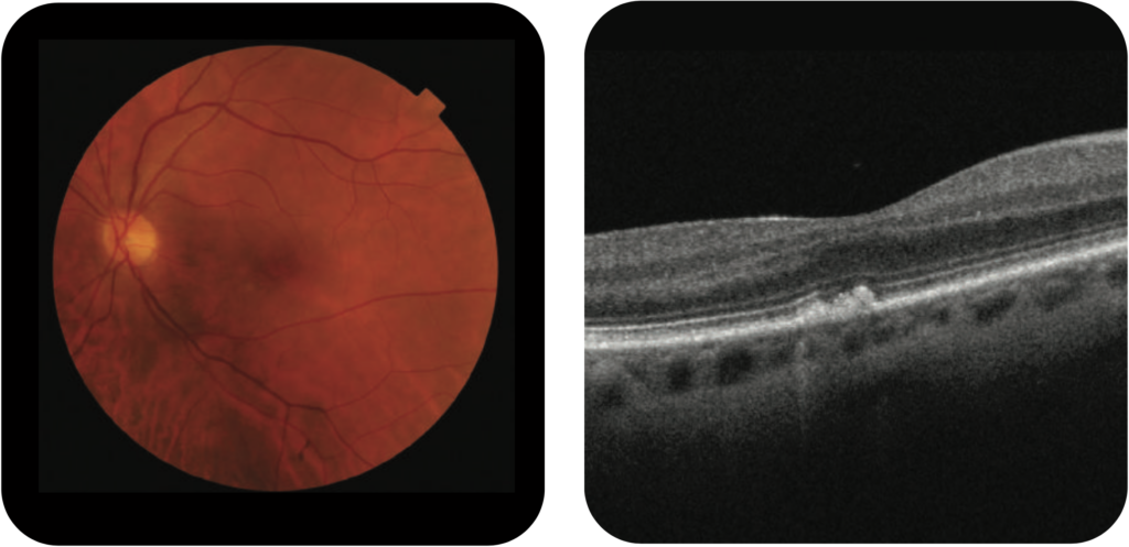
A retina may appear normal, but the detailed scan shows the irregularities caused by early AMD. AMD affects the macula which is the central part of the retina; the part where you have your best vision.
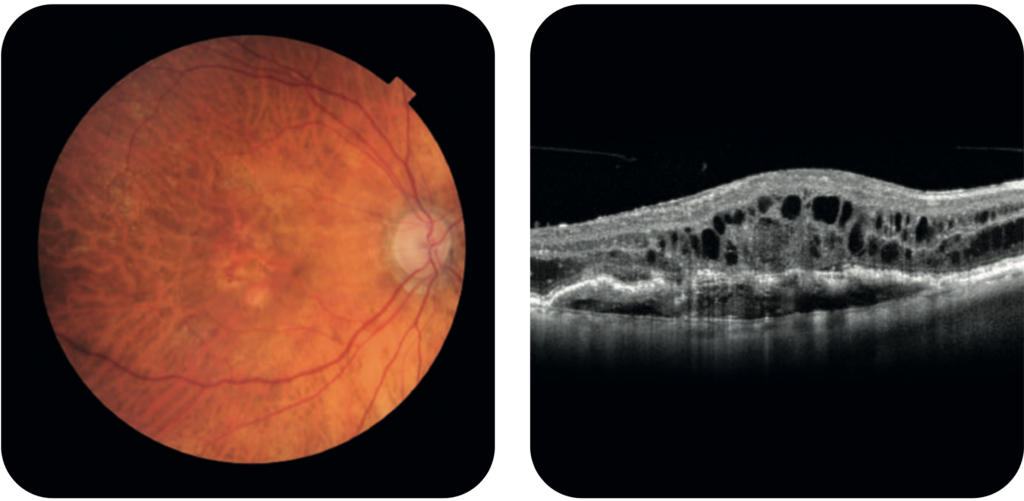
OCT scans are helpful in distinguishing what condition has caused a particular type of swelling.
Diabetic macular edema is swelling in the central retina caused by diabetes. OCT imaging is able to pick up the earliest stages so treatment can begin immediately.
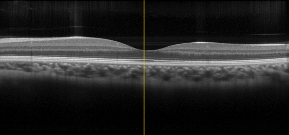
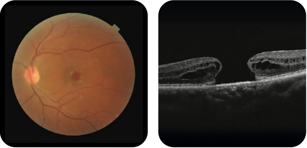
With small or early macular holes, things in your central vision will look blurry, wavy, or distorted. As the hole grows, a dark or blind spot appears in your central vision.
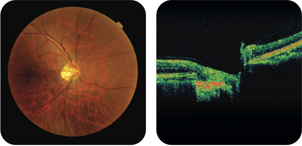
OCT provides lots of information about the optic nerve, which is the part of the eye affected by glaucoma. It is one of the best ways to help diagnose glaucoma as soon as possible and monitor changes to the optic nerve. OCT can be especially helpful in diagnosing forms of glaucoma that are not caused by an increase in eye pressure.
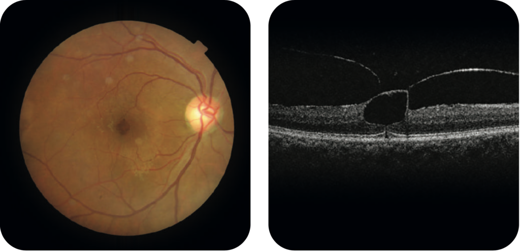
Vitreomacular traction is caused when the vitreous, a clear gel-like substance in the eye, pulls and tugs on the macula. This can damage the macula and cause vision loss if left untreated. We use OCT to help diagnose this condition.